Angiology and vascular surgery is a medical-surgical specialty dedicated to the study, prevention, clinical and instrumental diagnosis and treatment of vascular pathology.
Medical equipment
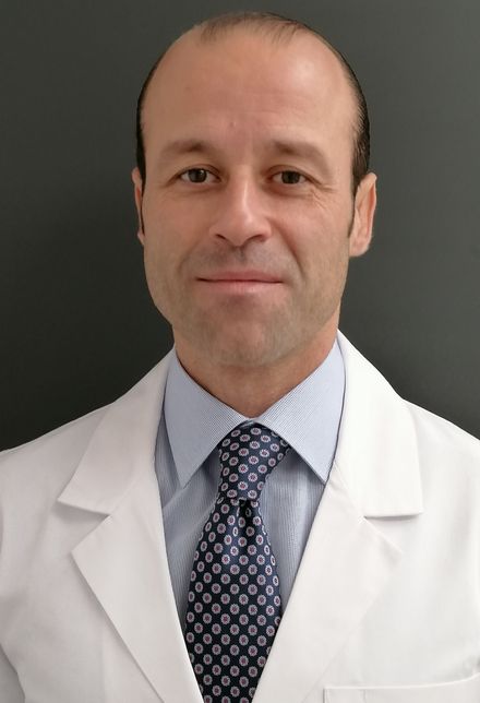
Luis Leiva
Head of Section
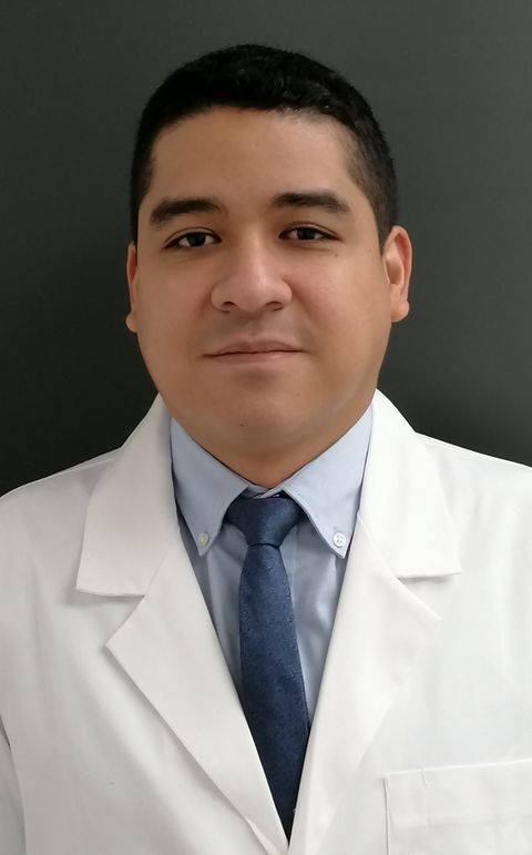
Juan Carlos Fletes Lacayo
Head of the Vascular Laboratory
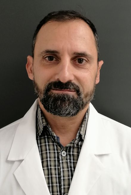
José Ignacio Fernandez Solares
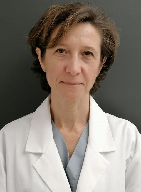
Ana Isabel Rodríguez Montalbán
VARICOSE VEINS
To understand the origin of varicose veins we must first know 2 aspects: the special anatomy of the venous system of the legs, and the functioning of the veins of the lower limbs that must fight against a natural force, gravity. Anatomy of the MMII venous system In the legs we find basically two types of veins: 1. Those that constitute the deep venous system, which usually accompanies the arteries and conducts 90% of the MMII blood to the heart. 2. And those that form the superficial venous system, which channels 10% of the blood of the MMII, and that forms a network below the skin, in which 2 veins, the internal saphenous vein and the external saphenous vein stand out . But these two systems are also intertwined by a set of connection veins called perforators. Although varicose veins are preferably dilations of the superficial network, they can be caused by a dysfunction in any of the three groups of veins. Operation of the leg veins The function of the leg veins is to collect and transport blood to the heart. Therefore, in the human being, when moving vertically, he must overcome the force of gravity. For this, the veins have 2 assistive mechanisms: the valves and the muscular pump. The valves are devices that are inside the veins, approximately every 10 centimeters, that act as floodgates, allowing the passage of blood only in one direction, from the feet to the heart. But on the other hand, when we walk, the leg muscles act as a pump, since their contraction and relaxation, squeezes the veins and empties them intermittently. What are varicose veins? Varicose veins are segmental dilations, more or less diffuse and more or less elongated, of superficial veins of the legs. Depending on its size and its distribution or origin, we can talk about several types: Truncal Varicose Veins. Collateral Varicose Veins. Reticular Varicose Veins. Telangiectasias or spider veins. The first 3 can be cataloged essentially as a medical problem, since they reflect a more severe Chronic Venous Insufficiency disorder. However, varicules and telangiectasias preferentially enter the field of Vascular Aesthetics. How are varicose veins removed? Before describing the possible therapeutic techniques, I would like to make some concepts clear: The most prepared specialist for the treatment of varicose veins is the Angiologist and Vascular Surgeon. Varicose veins have an important genetic load, and that makes some people relapse after an intervention. It is a mistake to think that with a single therapeutic option we will be able to eliminate any type of varicose vein. The vascular surgeon must master several techniques to offer the most appropriate for each case. Sometimes a patient may need several treatments to achieve complete elimination. In our group, after extensive experience with all possible techniques to treat varicose veins, we have opted to select 5 options, either as isolated techniques, or on many occasions, combined with each other: Liquid sclerotherapy and Microfoam Ambulatory Phlebectomy CHIVAL surgery Radiofrequency thermal Endoablation Mechanical-chemical Endoablation with Clarivein
Aortic aneurysm
Abdominal aortic aneurysm is a permanent and progressive dilation of the aortic artery mainly at the infrarenal level, which usually occurs in people over 50 years. Since this disease is usually asymptomatic, it can go unnoticed and its outcome can be fatal. At present, his diagnosis is based on an adequate directed medical history and complete physical examination, as well as a simple abdominal Doppler ultrasound carried out by a vascular surgeon. For many years, a lot of research has been done on the discovery of markers, usually protein, that allow establish a relationship with the development of the aortic aneurysm. Recently, researchers from the Center for Biomedical Research in Network of Cardiovascular Diseases (CIBERCV) (Spain), have managed to identify a protein whose elevation may be indicative of suffering from an abdominal aortic aneurysm, we talk about the CCL20 protein, whose levels are higher in patients with aortic aneurysm, than in healthy people or patients with non-aneurysmal atherosclerotic disease (patient with arterial irrigation insufficiency). This new finding is a great advance in the prevention and diagnosis of this disease, since it could allow an early diagnosis of the same. And although it is still under study, we can say that there is a new light for all those people who suffer from this pathology and who do not yet know it.
Intermittent claudication.
Arteriosclerosis is a disease characterized by the progressive narrowing of the arteries until their obstruction. As a result, there is a deficit in oxygen supply to the affected territories. One of the most characteristic are the legs, and in this case it is called Chronic Ischemia of MMII or Peripheral Arterial Disease. The prevalence of this disease in the Spanish population ranges between 2 and 10%. But there are a number of risk factors that can make this disease more frequent. We highlight several: age (especially above 60 years), male gender, smoking, diabetes mellitus, increased cholesterol, high blood pressure. We can distinguish 4 degrees of involvement: Grade I, where the arteries have begun to narrow but there are still no symptoms. It is difficult to detect these patients on time, but it is extremely important to begin with preventive measures and treatments as soon as possible. An effective measure is the review by a Vascular Surgeon of the patient who has risk factors, and if necessary, will perform a simple arterial EcoDoppler. Grade II, where the blood deficit determines that the leg muscles complain of pain when They are asked for some activity. It is this particular section that receives the name of Intermittent Claudication or Showcase Disease, since the patient is forced to stop his march at more or less constant distances. It is a characteristic pain usually located in the area of the twins of the leg, which usually develops after walking a certain distance, and that usually forces you to stop. The pain appears earlier if the patient climbs slopes or even stairs. In grade III, the lack of blood is so important that the pain appears even at rest, especially at the foot. The patient usually looks for a position where the feet are in decline or hanging, so that the gravity allows a greater contribution of blood. For this reason the patient usually says that it hurts more at night. And in grade IV, lesions and ulcers develop that can be predecessors of gangrene. Grade III and IV are known as Critical Ischemia, since the viability of the limb is seriously threatened, and the action by the Vascular Surgeon must be diligent. WHICH IS THE TREATMENT? The treatment includes several aspects. First of all, exercise is very important. Going for a walk every day is essential. Walking is the best stimulus for small arteries to develop and supply the narrowed or clogged artery. Other exercises or sports can be done, but the really important one is walking. Secondly it is crucial to control the risk factors. That is, we must apply the preventive and therapeutic measures necessary for the proper management of aspects such as cholesterol increase, high blood pressure and diabetes mellitus. In this sense, a correct multidisciplinary assessment and understanding between the attending physician and the Vascular Surgeon is important. But as the most important action to highlight in this section, in the case of smokers, it is to quit the smoking habit as soon as possible. The relationship of the appearance of atherosclerosis with tobacco is strongly demonstrated, but also its faster development and worse evolution if you do not quit smoking. Thirdly we must refer to the medication, which the safe vascular surgeon will best propose to the patient attending to his background. There is medication aimed at slowing the progression of the disease, such as antiplatelet agents (eg aspirin) or statins, and medication aimed at increasing the distance of claudication, such as pentoxifylline or cilostazol.And finally we must mention that the Surgeon Vascular nowadays has a series of very varied surgical techniques, to manage to unblock the arteries when necessary. From simple angioplasties or dilations, through springs or stents, to more complex operations such as bypass. The Vascular Surgeon is the specialist prepared to prevent, diagnose, and treat this disease correctly.
Cerebrovascular Disease
Stroke or ischemic stroke occurs as a result of the cessation or decrease of blood flow to a territory of the brain, either momentary or permanent. The most common disease is atherosclerosis; and one of its most characteristic clinical forms is the atheromatous plaque of the carotid artery bifurcation. It consists of a progressive narrowing of the artery as a result of a thickening of the arterial wall, generally in relation to the lipid or cholesterol deposit. The most common risk factors are age, male gender, hypertension, smoking, increased cholesterol and diabetes or blood sugar.In treatment, in addition to giving up the habit of smoking and eliminating or controlling factors At risk, treatment with antiplatelet agents such as aspirin / adiro and anti-cholesterol drugs such as statins is important. In some cases, especially in major narrows of the artery, we must be more aggressive. Some patients would benefit from operations such as carotid endarterectomy, where the artery is cleaned inside and a patch placed to enlarge its diameter. In other cases, it may be more appropriate to place a spring or stent inside to widen the artery.
Thromboembolic disease
This group includes a group of diseases that are characterized by high mortality, and we can group them into two aspects, venous thrombosis and pulmonary embolism. Three risk factors for thromboembolic disease have been considered for many years: endothelial lesion of the wall of the veins, stasis or blood stagnation and impaired coagulation. When, for whatever reason, a venous thrombus is formed, it initially consists of an aggregate of platelets that is surrounded by a fibrin mesh. When this initial thrombus becomes obstructing a vein, it can grow and expand, usually at first with little adhesiveness to the venous wall, and with the risk therefore of detaching and traveling through the bloodstream until it hits the lung; We know this picture as pulmonary embolism. MMII VENOUS THROMBOSIS At the level of the legs can be basically three differentiated pictures, which from greater to less serious are: Deep vein thrombosis. It consists of the formation of a clot or thrombus in the deep or internal veins of the limb, and is characterized by the more or less abrupt appearance of pain, swelling (edema), muscular tension (usually in the area of the twins) and a slightly bluish or dark coloration. This picture requires an early diagnosis by a vascular surgeon, who if necessary will perform a simple exploration with color EcoDoppler, to apply the therapeutic measures as soon as possible. It is easy to confuse deep vein thrombosis with other situations such as fibrillary rupture, intramuscular hemorrhage, Rupture of a knee cyst, arthritis, tendinitis, lymphedema.The delay in diagnosis and initiation of treatment can basically entail two risks: Pulmonary embolism (which in some cases can be fatal) and posttrombotic syndrome (which refers to set of signs and symptoms that appear in the limb chronically after a venous thrombosis, and that can become very severe: permanent swelling, skin lesions, ulcers, varicose veins, etc.). Superficial venous thrombosis. It consists of the formation of a clot or thrombus in the superficial or external veins of the limb, and is characterized by the appearance of an inflamed and painful cord, usually along the inner side of the leg and / or the thigh. It is a less serious entity than the previous one. Varicophlebitis. It refers to the formation of a clot or thrombus in a preexisting varicose vein. In many cases it is the reason that makes patients decide to have varicose veins. RISK AND TREATMENT FACTORS Some risk factors are well known and others not so much: age, obesity, immobility, having had a previous venous thrombosis, recent surgery, pregnancy, cancer, heart disease, infectious diseases, and the whole set of genetic alterations that we can call thrombophilia states. Today the treatment of venous thrombosis of MMII includes several facets. But first of all, highlight the importance of timely diagnosis. On the one hand we must mention anticoagulation, that is, the use of heparin drugs or oral anticoagulants, for a variable time, whose objective is to convert blood, to understand us, more liquid. On the other hand the elastic containment, if possible with therapeutic means of pharmacy or orthopedics, and postural measures. In some situations it may be advisable to use fibrinolytic drugs to dissolve the thrombus (require hospital admission) or surgical or endovascular techniques with the same objective. These options only make sense in some circumstances and especially in the first days after thrombosis, hence the importance of early diagnosis by a vascular surgeon. Once the acute phase is over, periodic control by the vascular surgeon is very important, to prevent and manage the development of post-thrombotic syndrome.
Lymphedema
In addition to the arteries and veins, there are other vascular ducts that are usually the forgotten ones: the lymphatic vessels. The lymphatic system has the function of collecting extracellular fluid; Therefore it has a drainage function. At the limb level we can distinguish lymphatic, microscopic capillaries around the cells, lymphatic collectors, characterized by the presence of valves that only allow one-way flow, and lymph nodes, grouped in ganglionic chains and constitute stations of the lymphatic circulation. Approximately 90% of the interstitial fluid is collected by the venous system, and 10% by the lymphatic capillaries. Lymph is a clear, clear and transparent liquid that comes from extracellular fluids, and it travels slowly. Dysfunction or slowing of the lymphatic system results in lymphedema, that is, edema or swelling due to accumulation of lymphatic fluid. And this can be primary, due to intrinsic or anatomical dysfunction, often congenital (very characteristic in MMII), or secondary, for example after surgery or radiotherapy (as usually occurs in an arm after mastectomy). Today the most widely used diagnostic test is isotopic lymphography, which is performed in Nuclear Medicine units. It consists of the injection under the skin of a radiopharmaceutical, to understand us, a marked substance that is capable of being detected in the gamma cameras, or nuclear medicine detection devices. TREATMENT Unfortunately, lymphedema does not often have a curative treatment; Many of the surgeries that have been developed have not been successful. So it is very important to focus therapeutic measures on improving the quality of life of patients and reducing complications. In addition to avoiding overweight and abundantly moisturizing the skin, we can use drugs that help in interstitial drainage function. But perhaps the most important section of the treatment and with better results, is the Manual Lymphatic Drainage or lymphatic drainage massages. Some Pressotherapy techniques may be useful. Postural measures and elastic containment with compression stockings or specific bandages are very important. There are certain physical exercises (kinesitherapy) that help improve muscle pump function and lymphatic drainage. Do not forget the importance of the assessment and monitoring by a Vascular Surgeon, who among other things will rule out that there is no associated pathology of the venous system, using a simple color EcoDoppler.
Diabetic foot
Diabetic patients can develop a wide variety of foot injuries. All these injuries are known as Diabetic Foot. It is a neuropathic and hyperglycemic-induced alteration (that is, the increase in blood sugar), in which there is usually a traumatic trigger, and ends up developing an injury or ulceration in the foot. We can talk about several levels of factors: Basic or initial factor. It is the disease of Diabetes. Primary factors. Diabetes usually generates three types of affectation in the feet: neuropathy (nerve injury that leads to a decrease or loss of sensation), microangiopathy (injury to small microscopic arteries) and macroangiopathy (narrowing and obstruction of arteries that lead to ischemia or failure. associated blood supply). Secondary factors. Whether hematological, immunological, articular or dermal, and triggers. That they can be intrinsic (due to bone deformity or limited mobility) or extrinsic (due to mechanical, thermal or chemical trauma). We can also talk about different types of lesions: Neuropathic ulcer, Neuropathic arthropathy, Neuroischemic ulcer and infected diabetic foot. PRACTICAL TIPS AND RECOMMENDATIONS We want to reflect here some tips that may be useful especially to prevent the appearance of foot injuries in diabetic patients. Daily inspection of the foot. Hand inspection of the inside of the footwear. Use suitable footwear. Change socks twice a day. Never walk without footwear. Never use hot water bags or electric pads. Do not rush nail cutting. Go to the podiatrist in case of ingrown toenails or calluses. Wash the feet with soap and water, with special attention to drying, especially between the fingers. Measure the temperature of the water with the elbow. Apply moisturizer after bath. Go to your doctor. in case of suspicious injuries. And assessment by Vascular Surgeon.
TREATMENTS:
Watch full video on YouTube


